-
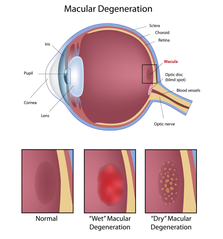
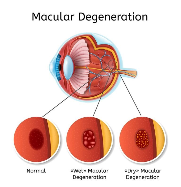
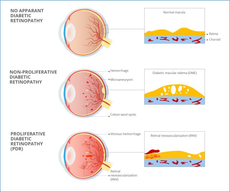
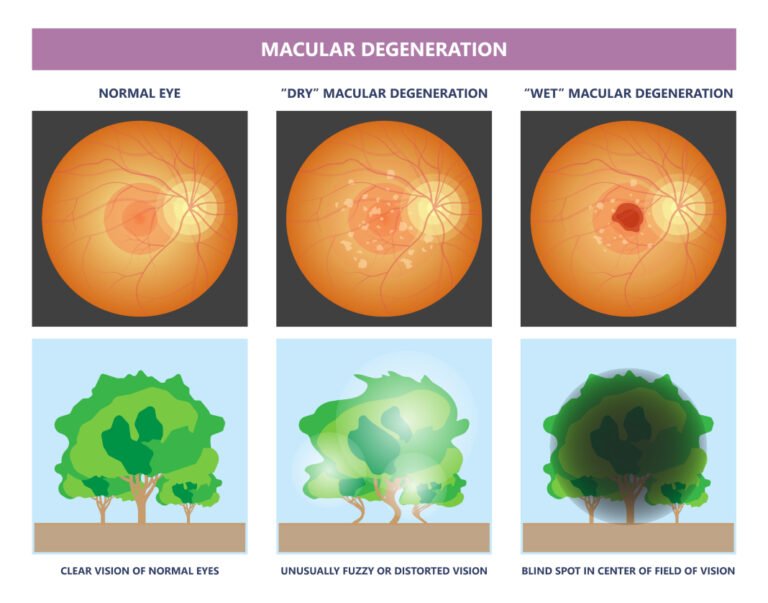
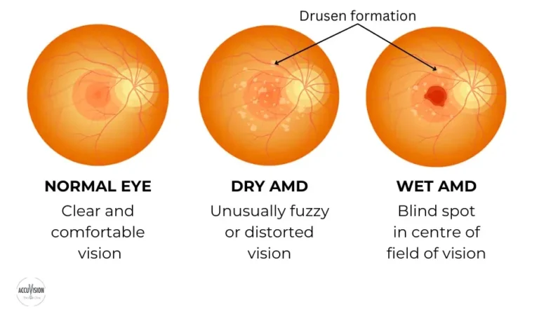
Types of AMD
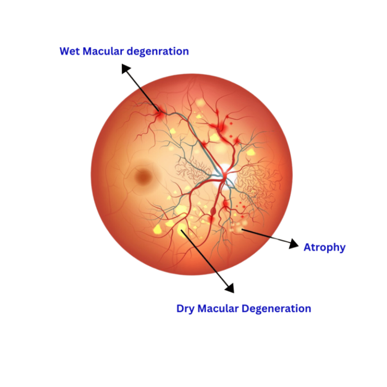
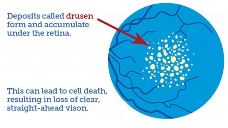
1. Dry AMD (Atrophic)
o Most common (90% of cases).
o Caused by thinning of the macula and accumulation of drusen (yellow
deposits).
o Progresses slowly over years.
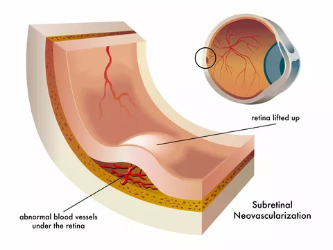
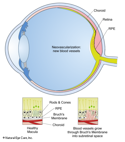
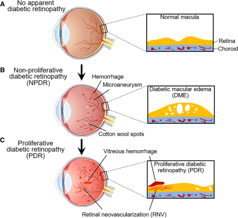
2. Wet AMD (Neovascular/Exudative)
o Less common but more severe.
o Caused by abnormal blood vessel growth under the retina, leading to
leakage & scarring.
Can cause rapid vision loss if untreated.
-
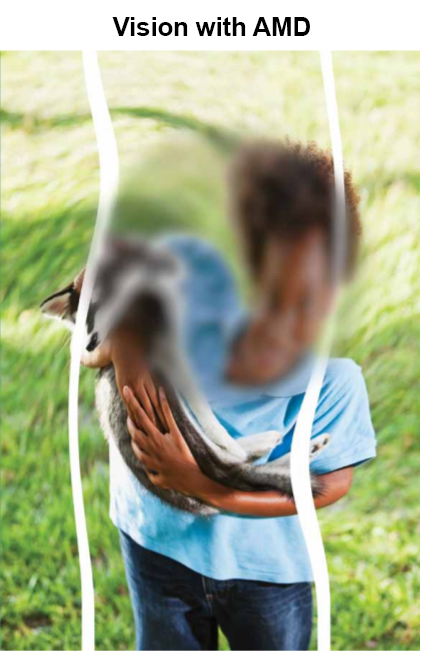
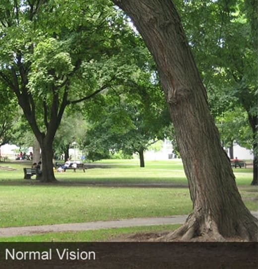
Symptoms of AMD
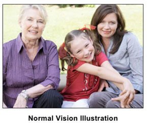
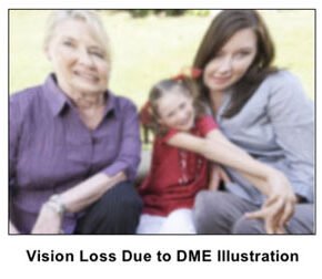
![]() Blurred or distorted central vision (e.g., straight lines appear wavy).
Blurred or distorted central vision (e.g., straight lines appear wavy).
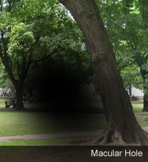
 Dark or empty spots in the center of vision.
Difficulty reading, recognizing faces, or seeing fine details.
Colors appear less bright.
Poor vision in low light.
Self-test: Use an Amsler Grid to check for vision distortions.
Causes & Risk Factors
• Aging (most common in people >50).
• Smoking (increases risk 3-4 times).
• Family history (genetics play a role).
• Cardiovascular diseases (high blood pressure, cholesterol).
• Treatment Options
1. Dry AMD
• No cure, but progression can be slowed with:
o AREDS2 supplements (Vitamin C, E, Zinc, Copper, Lutein, Zeaxanthin).
o Healthy diet (leafy greens, fish, nuts).
o Smoking cessation.
o Regular eye check-ups.
2. Wet AMD
• Anti-VEGF injections (Lucentis, Eylea, Avastin) to block abnormal blood vessels.
• Laser therapy (in some cases).
• Low-vision aids (magnifiers, screen readers).